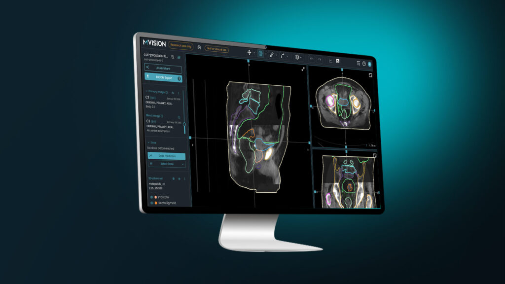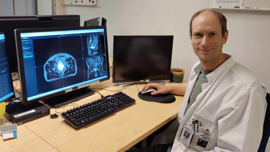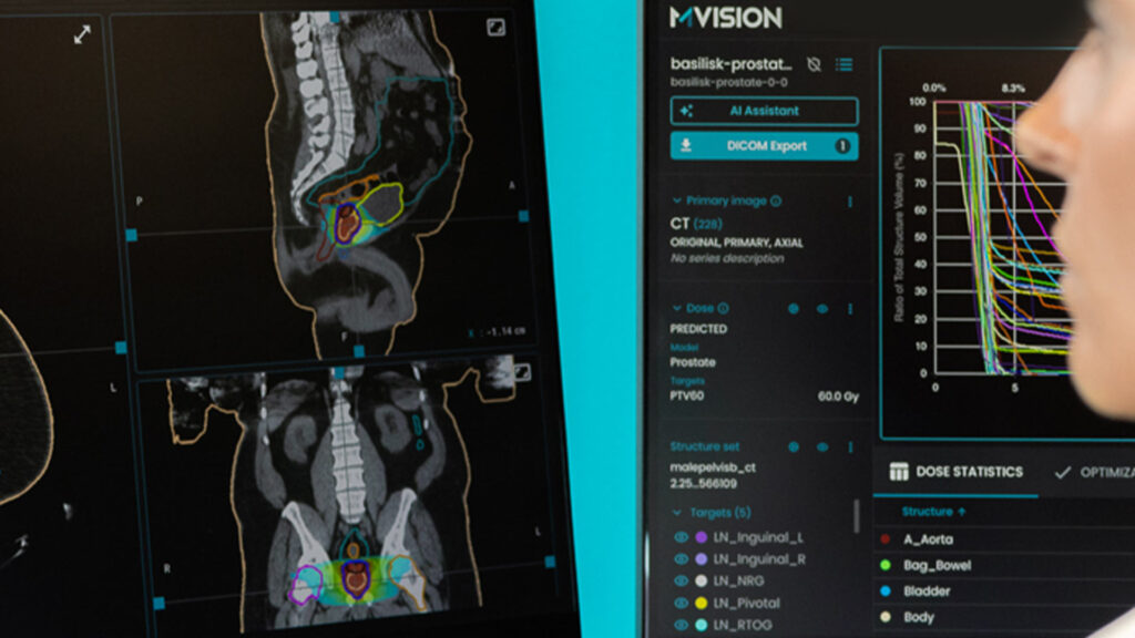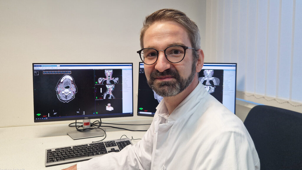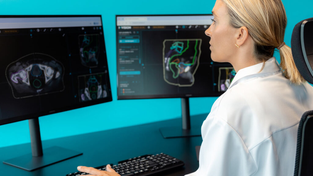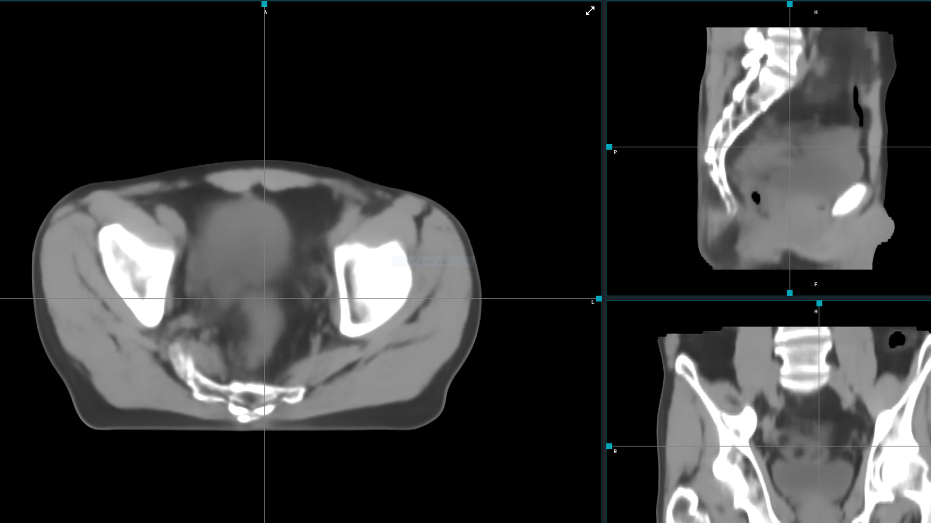
Delivering radiation therapy requires precise planning to obtain a certain dose of radiation in a specified volume within a patient’s body, while protecting the nearby tissues. The whole process relies on medical images (more frequently on CT scans), which are obtained using a certain protocol and dedicated setup devices. The workflow we use nowadays was developed in order to ensure reproducibility and accuracy. However, some patients already have had recent diagnostic CT, MRI or PET-CT scans when coming for radiation therapy. Acquiring additional CT images for treatment planning involves logistic issues, healthcare resources and an additional dose of ionizing radiation.
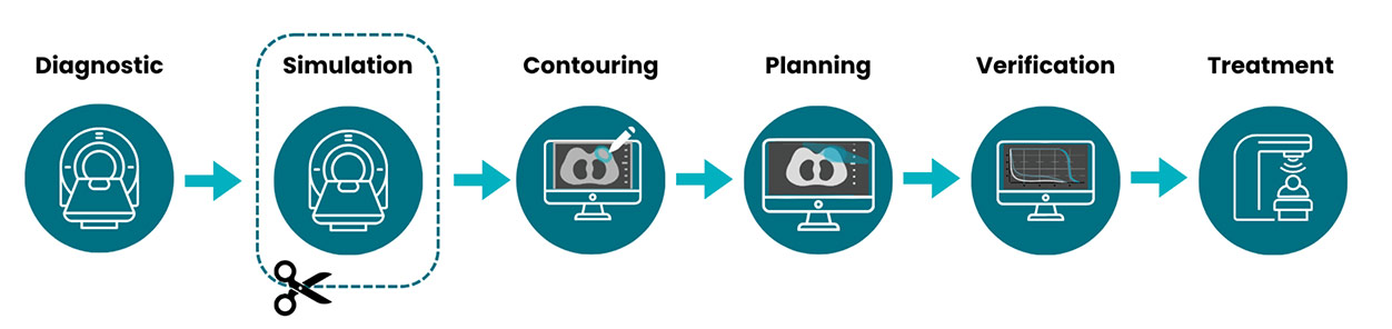
The ideal treatment workflow would reduce the number of steps and the irradiating diagnostic procedures and would speed up the process. Simulation-free planning represents such an opportunity. This alternative involves creating and delivering a radiotherapy plan from already-acquired diagnostic CT or MRI, rather than booking a dedicated CT simulation. The implementation case is strongest where the avoided appointment is a real access bottleneck and the diagnostic scan can be made geometrically and dosimetrically fit for purpose.
Expected advantages of Sim-free planning
Faster time to treatment and less patient burden matters most for urgent palliation, frail patients, and high-throughput pathways. A recent systematic review found diagnostic-CT planning shortens time to treatment, with reported low toxicity and effective symptom relief in the evaluated palliative settings [1].
Additionally, by removing one appointment, the scanner, therapist, contouring, and scheduling pressure is reduced. A prostate MRI-linac planning study explicitly frames sim-free radiation therapy as a route to lower treatment delays and improved cost-effectiveness [2].
Moreover, if a recent diagnostic study already shows target and relevant organs at risk at usable quality and suitable patient position, a second scan adds little clinical information while adding transfer, positioning, and waiting burden.
Feasibility of Sim-free planning
Practice changes need new ideas and documented proofs. Some reports can already be found on this topic, which are inspiring for new research projects.
- Diagnostic-CT based is currently the more operationally mature route for selected palliative cases. In one prospective implementation, all 30 patients were treated from diagnostic-CT plans; target-dose change after delivery was within −2% to +2.5%. PET/CT was particularly suitable, and pelvis/abdomen/thoracic or lumbar spine metastases were the most reproducible sites [3]. A 2025 review similarly reports diagnostic-CT dosimetric uncertainty under 5% in most cases, with thoracic/lumbar spine, sacrum, and pelvis as suitable single-fraction sites [1].
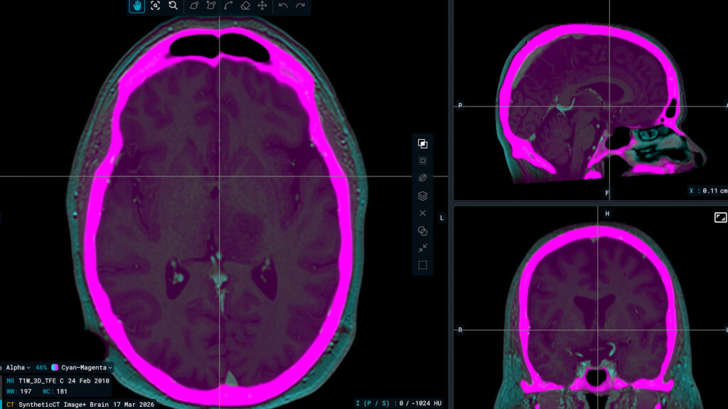
- MR-led/MR-only is most compelling where MRI improves target/OAR definition and adaptive MR-guided treatment supplies daily verification. In a small retrospective prostate MRI-linac comparison, simulation-free diagnostic-CT reference plans and adapted plans met institutional dose-volume criteria and had comparable plan quality to the standard workflow [4]. If MRI is also the sole dose-calculation source, a validated synthetic CT is required: one 10-patient prostate dosimetry study reported an approximately 1% clinically insignificant dose-distribution difference versus conventional CT [5].
| Where Image+ Fits In The feasibility question this article raises — can an already-acquired scan stand in for a dedicated simulation CT? — is the same question MVision AI's Image+ is built to answer. Image+ converts MRI, CBCT, or contrast-enhanced CT into synthetic CT images for photon dose calculation, and can enable radiotherapy planning preparation without dedicated CT acquisition. As with any sim-free workflow, clinicians remain in full control, reviewing all synthetic CT outputs before use. *Image+ is a module of Workspace+ which is a CE-marked (CE 2797) medical device under EU MDR 2017/745. Workspace+ is not available in all markets. |
Image and setup suitability
Since radiation therapy follows quality assurance and quality control rules, some elements need to be verified in order to keep the clinical standards.
For diagnostic CT, it is important to assess:
- field of view
- complete external contour
- slice thickness
- couch/immobilization and reproducible position
- HU integrity
- time since scan
- need for contrast/metal handling
CT evidence flags lung/low-density regions as more vulnerable to HU-related dose variation [3].
For diagnostic MRI, minimum requirements include a suitable MR sequence, large field of view, adequate slice resolution, and a commissioned synthetic-CT pathway if no CT is used. A planning study required a diagnostic 2D T1w large-FOV image covering all bony anatomy and a 2D T2w small-FOV image, excluding patients with hip implants. Scans with slice thickness up to 10.4 mm and FOV as small as 320 mm were usable after upsampling and body-contour extension, with dose calculated on a population-average bulk-density synthetic CT. In this cohort, 87.5% of patients had suitable diagnostic MRI, and an institutional audit found a similar proportion [2].
One legitimate concern involves setup uncertainties if no medical tattoos are available. Surface-guided radiation therapy could potentially compensate this need, so the departments that already have this technology available could implement more easily the sim-free approach.
Moreover, rectal and bladder filling might differ when treatment is delivered compared to diagnostic imaging. When using the traditional, sim-based approach, the variation is mitigated by using standardized protocols before simulation CT and before treatment. The same principle can be used for diagnostic imaging, if the radiology team is aware of these requirements.
Current and future perspectives
This new opportunity of sim-free planning can trigger workflow changes for both radiation therapy and radiology departments. Predefined imaging acquisition and acceptance rules can turn selected cases into an expedited pathway rather than an ad-hoc exception.
Some situations still need extra caution, not being suitable for this sim-free approach. Targets requiring steep dose gradients, major setup mismatch, or cases needing highly precise heterogeneity/dose modeling represent such cases in which a dedicated simulation scan is still needed [1].
However, a selective, protocolized replacement for CT simulation, not a universal removal of simulation, seems reasonable, especially when these changes improve the workflow. The curative MR-linac and MR-only are still in an early phase and disease-specific, but things will probably evolve in the future [4,5].
References
- Zwahlen et al., 2025. Simulation-free palliative radiotherapy using diagnostic CT scans– is less more? A systematic review. Clinical and Translational Radiation Oncology.
- Chick et al., 2025. Towards rapid and efficient simulation-free radiotherapy: MR guided adaptive prostate radiotherapy on the MR-Linac using diagnostic MRI reference planning. Radiotherapy and Oncology.
- Wong et al., 2020. Diagnostic CT enabled planning for palliative radiotherapy: removing the need for a planning CT. Practical Radiation Oncology.
- Warda et al., 2024. Simulation-free magnetic resonance-guided radiation therapy of prostate cancer. Physics and Imaging in Radiation Oncology.
- Benayun, 2023. Dosimetry Validation of MR-Only Treatment Planning Based upon the Synthetic MRCAT Pelvis with Continuous Hounsfield Units in Prostate Cancer. International Journal of Radiation Oncology, Biology, Physics.