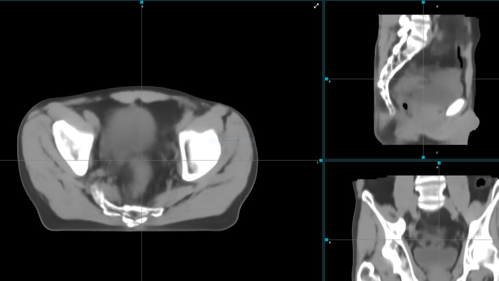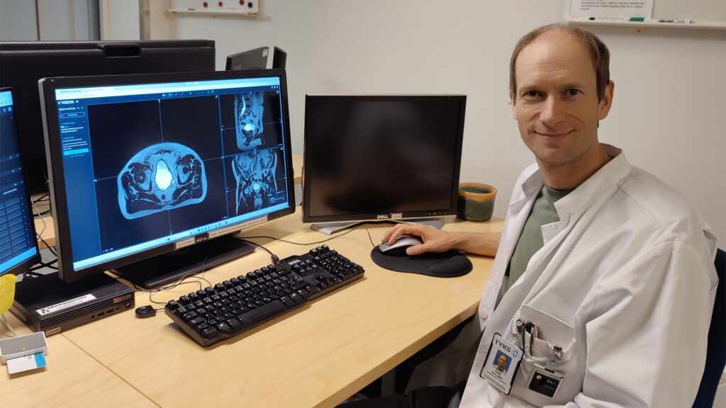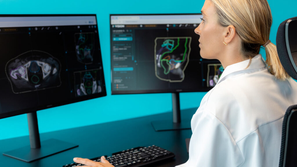
The evolution of radiotherapy is increasingly defined by the integration of advanced imaging modalities that transcend the limitations of conventional Computed Tomography (CT). While CT remains the gold standard for electron density mapping and dose calculation, its reliance on ionizing radiation and inferior soft-tissue contrast often complicates the clinical pathway. Modern workflows are now shifting toward MR-only planning, CBCT-based adaptive support, and automated contrast removal through the generation of synthetic CT (sCT) images. By utilizing deep learning and voxel-based algorithms, these techniques eliminate registration errors between disparate imaging sets, reduce cumulative radiation exposure, and streamline the patient journey from diagnosis to delivery. This shift not only improves geometric and dosimetric accuracy but also facilitates real-time adaptive interventions, ensuring that treatment remains highly conformal to the patient’s daily anatomy.
MR-only planning
MR-only planning workflows bring several key benefits to radiotherapy by eliminating registration errors, simplifying the patient pathway, reducing radiation exposure, and improving target delineation through superior soft-tissue contrast (1,2). MRI’s superior soft-tissue contrast enables more accurate identification of tumor boundaries and organs at risk compared to CT alone (1,3). This improved visualization can enable margin reduction, dose escalation to targets, and better sparing of critical structures.
However, the conventional radiotherapy workflow requires both CT simulation and MRI for dose calculation. Co-registration of diagnostic MRI and planning CT introduces systematic geometric errors. By implementing an MR-only workflow using synthetic CT (sCT) generation from MRI, this registration uncertainty is eliminated, potentially improving the overall geometric accuracy (1).
MR-only planning reduces the patient pathway from two separate imaging sessions to a single MRI acquisition, improving patient comfort and convenience (2). Recent studies demonstrate that simulation-free workflows using diagnostic MRI can save multiple weeks in the treatment pathway. One of those studies analyzed patients from the HERMES trial. Previously acquired diagnostic MR images were used to create a reference treatment plan without clinician input, using MVision Contour+®. Adapted plans were simulated based on those contours and compared with those from the traditional workflow.
Almost 9 in 10 patients had suitable diagnostic scans for reference planning. Online treatment plans were clinically acceptable for target dose and conformality. They met all mandatory clinical goals with no detriment to OAR dose or plan deliverability. Accuracy of the synthetic CT approach was high with gamma results at 2mm/2% all above 98.9% (4). The streamlined workflow also reduces staffing requirements and scheduling complexity (5).
Advanced atlas-based and deep learning voxel-based methods achieve dosimetric differences of less than 1% between sCT and actual CT planning, with positional verification deviations under 1 mm (1).
Studies report gamma pass rates exceeding 98% for photon plans and 97% for proton plans at clinically relevant criteria (6). For prostate radiotherapy specifically, the systematic difference in patient positioning using MR-only workflows averages less than 0.5 mm across all directions (7).
By eliminating the planning CT scan, radiation exposure is reduced, which is particularly valuable for patients requiring multiple treatment courses or adaptive replanning (1). This advantage extends to pediatric patients and those with concerns about cumulative radiation dose.
Adaptive support
CBCTs are useful for position verification and for adaptive radiotherapy, capturing real-time anatomical changes. However, CBCT has inaccurate Hounsfield unit (HU) values, artifacts, and noise that prevent direct use for dose calculation (8).
sCT images with reduced artifacts and improved soft-tissue contrast enable more accurate automated organ delineation compared to raw CBCT. A study on images from forty rectal cancer patients treated with IMR showed a mean absolute error reduction from 135.84 ± 41.59 HU for the CT and CBCT comparison to 52.99 ± 12.09 HU for the CT and sCT comparison (9).
For prostate cancer, Dice similarity coefficients for bladder and rectum reached 0.92 and 0.84 respectively on sCT, compared to 0.90 and 0.83 on raw CBCT (10). This improved segmentation accuracy reduces manual contouring time and supports faster adaptive replanning decisions.
Deep learning-based sCT generation corrects CBCT’s limitations, reducing mean absolute error from 69-131 HU for raw CBCT to 24-50 HU for sCT across multiple anatomical sites (11,12).
This HU accuracy enables reliable dose calculations with gamma pass rates exceeding 96-99% at clinically relevant criteria (2%/2mm or 1%/1mm), comparable to planning CT (12).
Several studies demonstrated that sCT better captures interfractional variations in organ filling, tumor regression, and weight loss throughout treatment. For a series of thirty patients treated for pancreatic cancer, mean absolute error was lower in HU for sCT compared to the original raw CBCT. Additionally, statistically significant differences (P < 0.05) were found between the CT- and the CBCT-based plans. By contrast, no significant differences (P > 0.05) were observed in the PTV and OAR dose-volume-histogram (DVH) metrics between the CT- and sCT-based plans (13).
Applicability to both photon and proton therapy extends the clinical utility. sCT generation has demonstrated high dosimetric accuracy for proton therapy, which is particularly sensitive to HU errors due to range uncertainties. Studies show gamma pass rates of 97-99% for proton plans and water equivalent thickness differences of only 1.3-1.9 mm, supporting daily proton dose verification and adaptation (14,15).
CBCT-based sCT enables clinicians to assess whether anatomical changes warrant plan adaptation by calculating actual delivered doses rather than relying on geometric changes alone. Studies demonstrate that online adaptive radiotherapy using CBCT-based sCT significantly improves target coverage (CTV D98% from 97.85% to 98.55%) and plan acceptability (from 24.8% to 98%) compared to non-adaptive approaches (16).
CBCT-based sCT leverages imaging already acquired daily for positioning, transforming it into a planning-quality dataset without additional scans. This approach has been successfully implemented across multiple disease sites including head-and-neck, pelvis, thorax, and breast cancer (12).
Reduced treatment time compared to MRI-guided workflows makes CBCT-based adaptation more practical for routine clinical use. While maintaining comparable dosimetric accuracy to MRI-based synthetic CT approaches, CBCT acquisition is faster and more readily available on conventional linear accelerators.
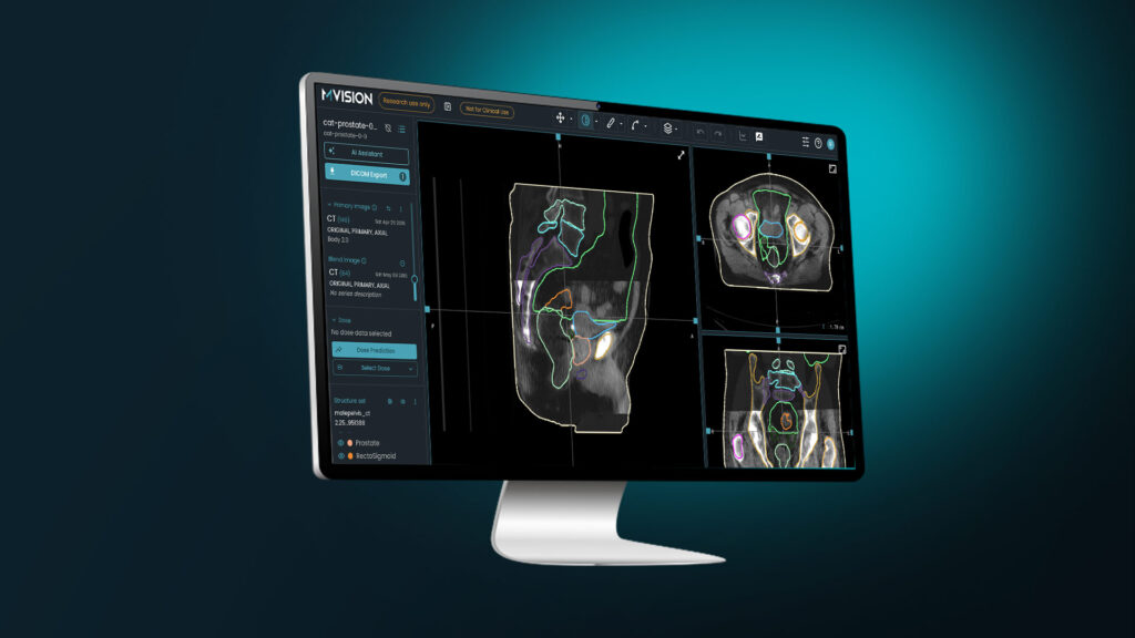
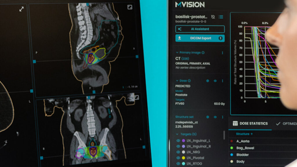
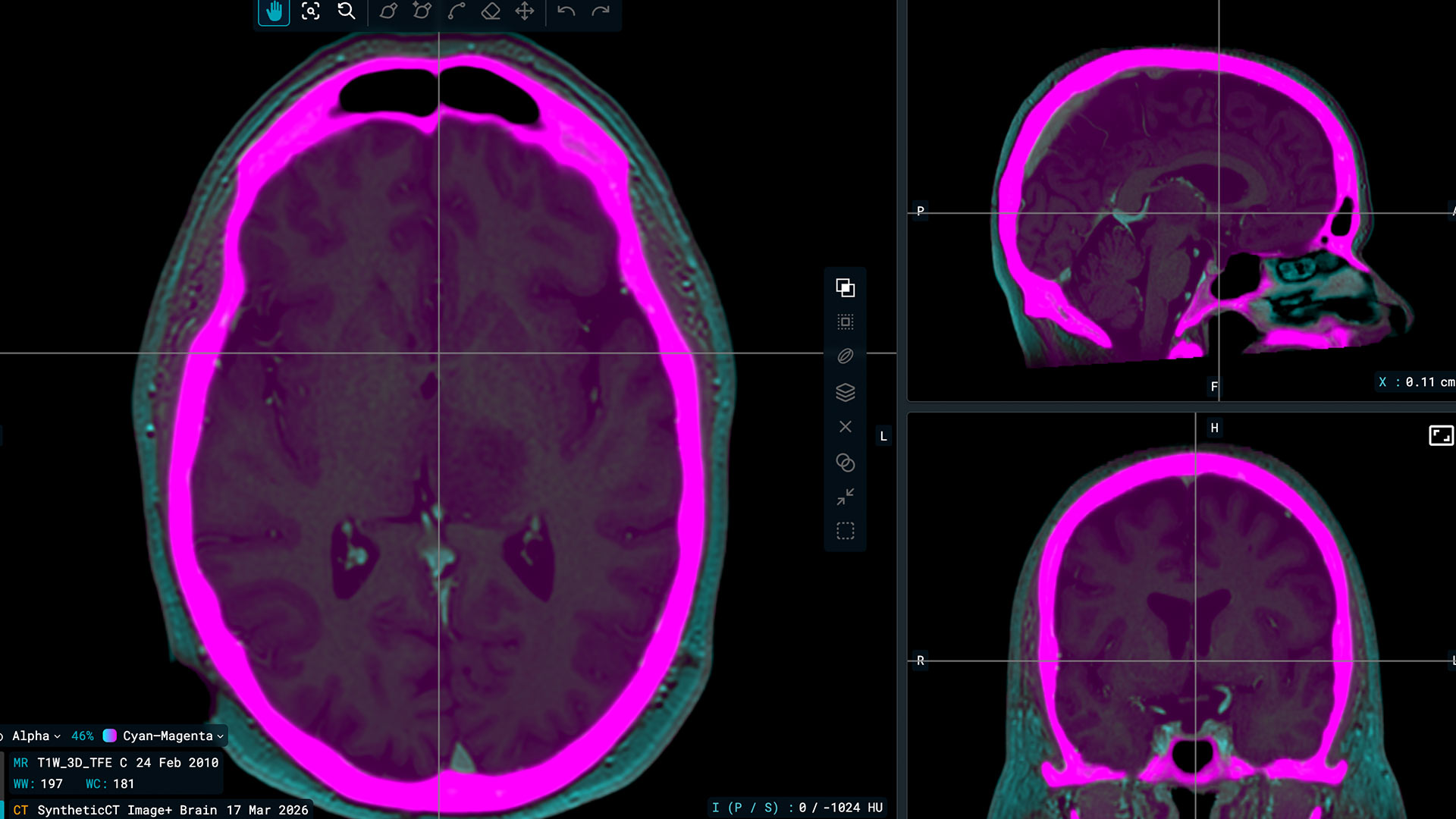
Image+, MVision AI’s synthetic imaging module within the Workspace+ platform, is designed to support radiotherapy preparation workflows. Image+ converts MRI, CBCT, and contrast-enhanced CT scans into synthetic CT images to support photon dose calculation in treatment preparation and offline adaptive workflows. Clinicians review all synthetic CT outputs before use to confirm alignment with institutional clinical standards and applicable verification procedures. The module supports MR-only workflows, CBCT-based dose recalculation, and virtual non-contrast image generation using imaging systems already available in the clinic.
Contrast removal
Contrast enhancement is often recommended for superior target delineation, particularly for head and neck, lung, and abdominal malignancies (17,18). However, iodinated contrast agents significantly increase HU values due to iodine’s high atomic number. Treatment planning systems erroneously interpret these elevated HU values as high-density tissue, leading to overestimation of beam attenuation (19).
Phantom studies demonstrated that contrast enhancement can increase HU values by 145-274 HU, potentially causing dose calculation errors of 2.3-7.4% for photon beams depending on contrast concentration and beam energy (19). The magnitude of error is particularly problematic for proton therapy, where conventional contrast-enhanced CT can produce dose errors exceeding 3.6% and range errors up to 3.2 mm due to incorrect stopping power calculations (20,21).
For whole-brain, whole-neck, mediastinal, and whole-pelvic irradiation, mean increases in monitor units are typically less than 1% and considered negligible. However, upper abdominal radiotherapy shows mean dose increases exceeding 2%, making this region particularly susceptible to contrast-related errors (22). The impact depends on the volume and concentration of contrast within the treatment field.
Standard practice traditionally requires two separate CT acquisitions: a non-contrast scan for dose calculation and a contrast-enhanced scan for target delineation, which are then co-registered. The NCCN guidelines for lung cancer recommend IV contrast for better target and organ delineation in patients with central tumors or nodal disease, but note that “density masking or use of a pre-contrast scan may be needed when intense enhancement is present” (17). Similarly, ASTRO consensus guidelines recommend using contrast-enhanced scans to assist with tissue delineation but acknowledge that “large areas of contrast may need a density override or a registered non-contrast scan for planning purposes if volumes of contrast significantly influence dose calculations” (23).
Deep learning methods can generate synthetic non-contrast images from conventional contrast-enhanced CT, achieving mean absolute errors of 6.7 HU and gamma pass rates of 99.6% (24).
In conclusion, the implementation of synthetic CT technology represents a transformative leap in radiotherapy, offering a robust solution to the long-standing challenges of image co-registration and dose calculation inaccuracies. Whether by enabling an MR-only workflow that simplifies the patient pathway, leveraging CBCT for precise daily adaptive replanning, or neutralizing the dosimetric interference of iodinated contrast agents, sCT ensures that high-quality, planning-grade data is available at every stage. With gamma pass rates consistently exceeding 97-99% across both photon and proton therapies, these AI-driven methods provide a level of accuracy comparable to traditional CT simulation while significantly reducing manual labor and treatment delays. Ultimately, these advancements pave the way for a more efficient, patient-centric, and highly personalized approach to cancer treatment.
References
- Johnstone E, Wyatt JJ, Henry AM, et al. Systematic Review of Synthetic Computed Tomography Generation Methodologies for Use in Magnetic Resonance Imaging-Only Radiation Therapy. Int J Radiat Oncol Biol Phys. 2018;100(1):199-217. doi:10.1016/j.ijrobp.2017.08.043
- Kerkmeijer LGW, Maspero M, Meijer GJ, van der Voort van Zyp JRN, de Boer HCJ, van den Berg CAT. Magnetic Resonance Imaging only Workflow for Radiotherapy Simulation and Planning in Prostate Cancer. Clin Oncol (R Coll Radiol). 2018;30(11):692-701. doi:10.1016/j.clon.2018.08.009
- Owrangi AM, Greer PB, Glide-Hurst CK. MRI-only treatment planning: benefits and challenges. Phys Med Biol. 2018;63(5):05TR01. Published 2018 Feb 26. doi:10.1088/1361-6560/aaaca4
- Chick J, Casey F, Cooper S, et al. Towards rapid and efficient simulation-free radiotherapy: MR guided adaptive prostate radiotherapy on the MR-Linac using diagnostic MRI reference planning. Radiother Oncol. 2025;211:111053. doi:10.1016/j.radonc.2025.111053
- Rong Y, Tegtmeier R, Clouser EL Jr, et al. Advancements in Radiation Therapy Treatment Workflows for Precision Medicine: A Review and Forward Looking. Int J Radiat Oncol Biol Phys. 2025;122(4):1022-1034. doi:10.1016/j.ijrobp.2025.04.010
- Huijben EMC, Terpstra ML, Galapon AJ, et al. Generating synthetic computed tomography for radiotherapy: SynthRAD2023 challenge report. Med Image Anal. 2024;97:103276. doi:10.1016/j.media.2024.103276
- Kemppainen R, Vaara T, Joensuu T, Kiljunen T. Accuracy and precision of patient positioning for pelvic MR-only radiation therapy using digitally reconstructed radiographs. Phys Med Biol. 2018;63(5):055009. Published 2018 Mar 2. doi:10.1088/1361-6560/aaad21
- Chen L, Liang X, Shen C, Nguyen D, Jiang S, Wang J. Synthetic CT generation from CBCT images via unsupervised deep learning. Phys Med Biol. 2021;66(11):10.1088/1361-6560/ac01b6. Published 2021 May 31. doi:10.1088/1361-6560/ac01b6
- Zhao J, Chen Z, Wang J, et al. MV CBCT-Based Synthetic CT Generation Using a Deep Learning Method for Rectal Cancer Adaptive Radiotherapy. Front Oncol. 2021;11:655325. Published 2021 May 31. doi:10.3389/fonc.2021.655325
- Chen X, Liu Y, Yang B, et al. A more effective CT synthesizer using transformers for cone-beam CT-guided adaptive radiotherapy. Front Oncol. 2022;12:988800. Published 2022 Aug 25. doi:10.3389/fonc.2022.988800
- Yeap PL, Du X, Zhou M, Hoole A, Barnett GC, Jena R. Few-shot CBCT-based synthetic CT generation with denoising diffusion probabilistic model. Med Phys. 2025;52(11):e70126. doi:10.1002/mp.70126
- Prunaretty J, Colombo L, Romdhani S, et al. Self-learning GAN based synthetic CT generation: unlocking CBCT-based adaptive radiotherapy. Front Oncol. 2026;16:1756153. Published 2026 Feb 20. doi:10.3389/fonc.2026.1756153
- Liu Y, Lei Y, Wang T, et al. CBCT-based synthetic CT generation using deep-attention cycleGAN for pancreatic adaptive radiotherapy. Med Phys. 2020;47(6):2472-2483. doi:10.1002/mp.14121
- Pang B, Si H, Liu M, et al. Comparison and evaluation of different deep learning models of synthetic CT generation from CBCT for nasopharynx cancer adaptive proton therapy. Med Phys. 2023;50(11):6920-6930. doi:10.1002/mp.16777
- Vestergaard CD, Muren LP, Elstrøm UV, et al. Daily proton dose re-calculation on deep-learning corrected cone-beam computed tomography scans. Radiother Oncol. 2025;209:110953. doi:10.1016/j.radonc.2025.110953
- Vestergaard CD, Muren LP, Elstrøm UV, et al. Daily proton dose re-calculation on deep-learning corrected cone-beam computed tomography scans. Radiother Oncol. 2025;209:110953. doi:10.1016/j.radonc.2025.110953
- National Comprehensive Cancer Network. NCCN clinical practice guidelines in oncology (NCCN guidelines®): non-small cell lung cancer. Version 2.2026 [Internet]. Plymouth Meeting (PA): National Comprehensive Cancer Network; 2026 Mar 13 [cited 2026 Apr 9]. Available from: https://www.nccn.org/guidelines/category_1
- National Comprehensive Cancer Network. NCCN clinical practice guidelines in oncology (NCCN guidelines®): Head and Neck Cancers. Version 2.2026 [Internet]. Plymouth Meeting (PA): National Comprehensive Cancer Network; 2026 Mar 13 [cited 2026 Apr 9]. Available from: https://www.nccn.org/guidelines/category_1
- Ramm U, Damrau M, Mose S, Manegold KH, Rahl CG, Böttcher HD. Influence of CT contrast agents on dose calculations in a 3D treatment planning system. Phys Med Biol. 2001;46(10):2631-2635. doi:10.1088/0031-9155/46/10/308
- Ates O, Hua CH, Zhao L, et al. Feasibility of using post-contrast dual-energy CT for pediatric radiation treatment planning and dose calculation. Br J Radiol. 2021;94(1118):20200170. doi:10.1259/bjr.20200170
- Lalonde A, Xie Y, Burgdorf B, et al. Influence of intravenous contrast agent on dose calculation in proton therapy using dual energy CT. Phys Med Biol. 2019;64(12):125024. Published 2019 Jun 21. doi:10.1088/1361-6560/ab1e9d
- Shibamoto Y, Naruse A, Fukuma H, Ayakawa S, Sugie C, Tomita N. Influence of contrast materials on dose calculation in radiotherapy planning using computed tomography for tumors at various anatomical regions: a prospective study. Radiother Oncol. 2007;84(1):52-55. doi:10.1016/j.radonc.2007.05.015
- Wright J, Yom S, Awan M . et al. Standardizing Normal Tissue Contouring for Radiation Therapy Treatment Planning: An ASTRO Consensus Paper Practical Radiation Oncology, 2018; 9, 65-72
- Liugang G, Kai X, Chunying L, et al. Generation of Virtual Non-Contrast CT From Intravenous Enhanced CT in Radiotherapy Using Convolutional Neural Networks. Front Oncol. 2020;10:1715. Published 2020 Sep 8. doi:10.3389/fonc.2020.01715
*CE-marked (CE 2797) medical device under EU MDR 2017/745; Workspace+ not available in all markets.