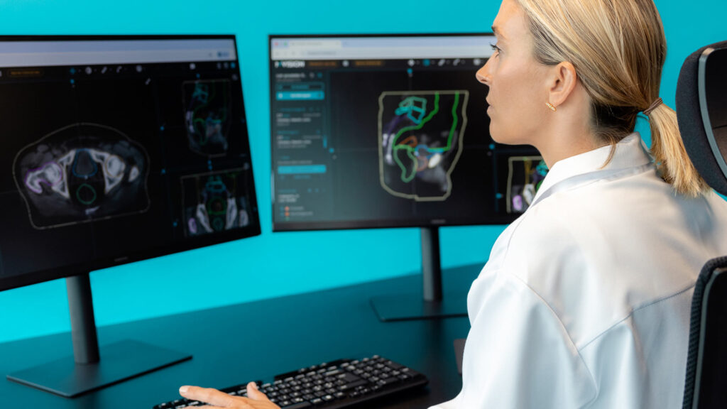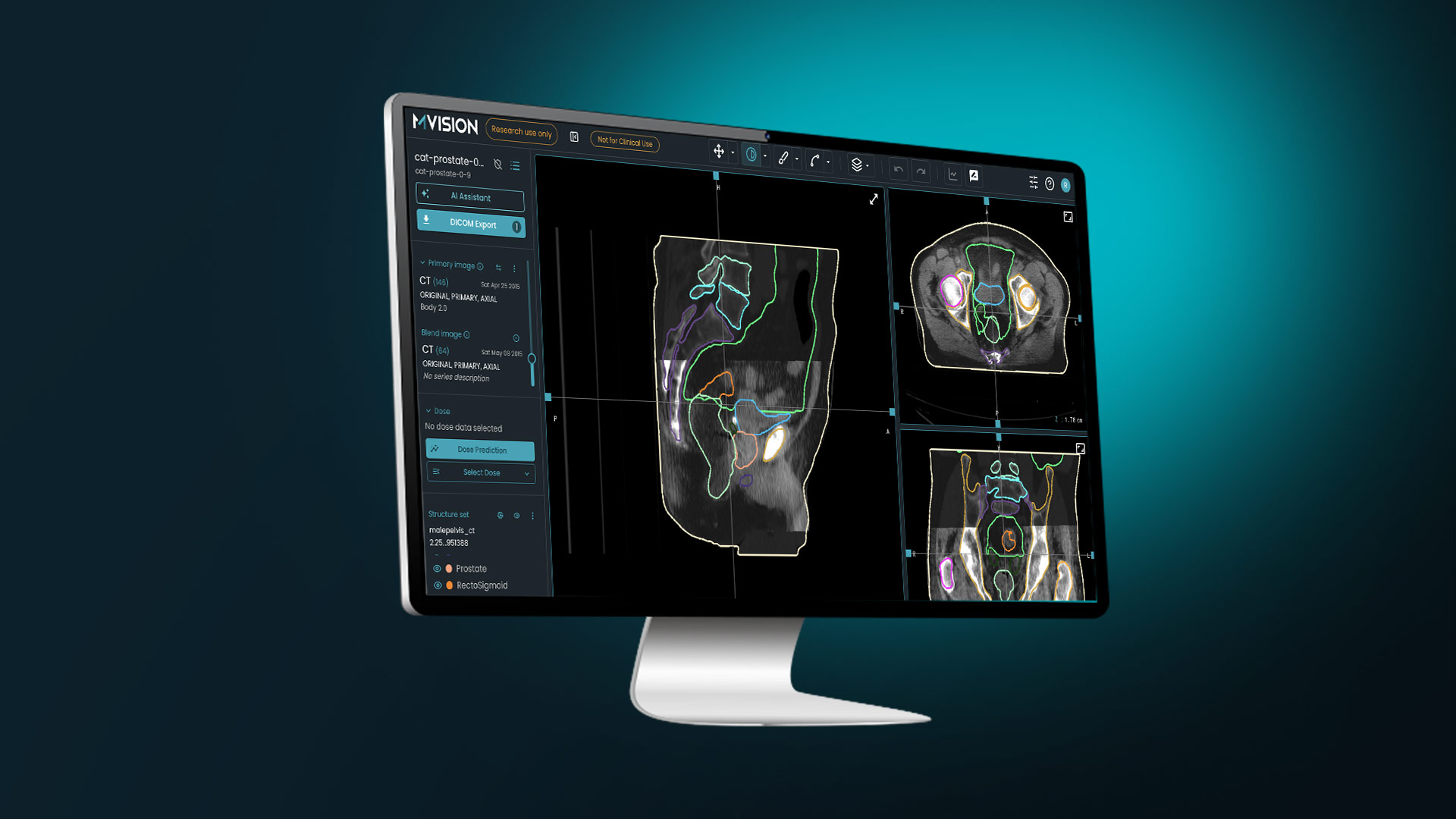
In modern radiation oncology, adaptive radiotherapy (ART) addresses dynamic anatomical changes that occur throughout a multi-week course of treatment (Dona Lemus et al., 2024). These changes—such as tumor shrinkage, weight loss, or shifts in internal organ filling—can degrade target coverage and inadvertently increase radiation exposure to surrounding organs at risk (OARs).
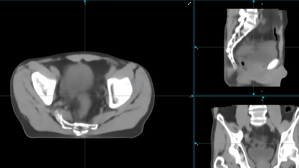
While online ART performs re-planning with the patient on the treatment couch, offline ART evaluates cumulated doses and re-optimizes treatment plans asynchronously between treatment fractions. Central to the offline ART workflow is contour propagation: the automated mapping of target volumes and OAR contours from a baseline planning computed tomography (pCT) scan onto subsequent fractional images (repeat CTs or on-treatment cone-beam CTs).
1. Practical Implementation
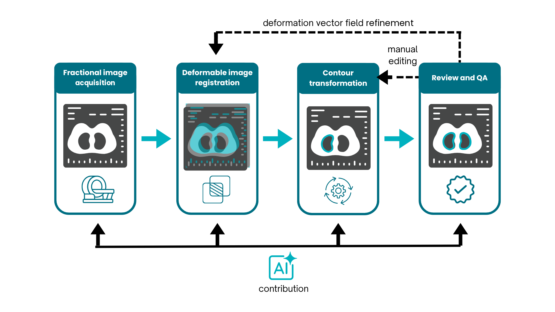
The practical clinical implementation of contour propagation in offline ART generally follows a structured multi-step pipeline:
- Fractional Image Acquisition: During or between treatment fractions, repeat 3D imaging—such as a re-simulation CT or on-board Cone-Beam CT (CBCT)—is acquired to capture the patient’s updated anatomy.
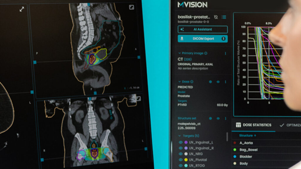
- Deformable Image Registration (DIR): A registration algorithm establishes spatial correspondences between voxels in the reference pCT and the fractional image, computing a 3D Deformation Vector Field (DVF) (Rong et al., 2021). Common algorithm families include intensity-based optical flow, B-spline free-form deformations, Demons algorithms, and emerging patient-specific deep learning (DL) neural networks (Li et al., 2017; Smolders et al., 2023).
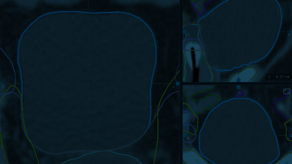
- Contour Transformation: The computed DVF is applied to the original physician-delineated structures on the pCT, mathematically mapping their boundaries onto the new fractional image space.
- Physician Review and Quality Assurance (QA): Because automated algorithms are subject to spatial uncertainties, clinicians inspect and manually edit propagated contours before using them for dose re-calculation or plan re-optimization (Hardcastle et al., 2013). Advanced techniques, such as contour-guided DIR (CG-DIR), can re-incorporate these clinician edits to iteratively refine the underlying DVF (Gu et al., 2013).
2. Advantages and Challenges
Offline adaptive radiotherapy improves workflow efficiency by reducing the time that would have been needed for manual re-contouring. It also enables accurate dose tracking and accumulation across the entire treatment course for target volumes and OARs. As a result, the dose coverage is preserved but the dose to the OARs is reduced (Cagni et al., 2022)(Hardcastle et al., 2013).
Despite its benefits, contour propagation in offline ART faces several critical technical and clinical limitations:
- Sensitivity to Image Quality Disparities: CBCT images often suffer from scatter artifacts, lower soft-tissue contrast, and Hounsfield Unit (HU) inaccuracies compared to diagnostic planning CTs. These discrepancies hamper intensity-based DIR algorithms, leading to larger contour errors in low-contrast soft tissues like esophagus or nodal targets (Li et al., 2016; Li et al., 2017).
- Non-Physical Deformations and Disappearing Masses: Classical DIR algorithms assume mass conservation. When tumors shrink significantly or cavities fill with fluid, mathematical registration algorithms may generate unphysical deformation vector fields, warping target boundaries incorrectly (Rong et al., 2021).
- Dependence on Quality Assurance: Unedited propagated contours cannot be used blindly. Errors in propagated OAR or target boundaries can lead to systematic under-dosing of targets or over-dosing of critical structures if used directly in re-optimization (Hardcastle et al., 2013). Routine implementation requires rigorous QA protocols guided by standard professional benchmarks, such as AAPM Task Group 132 recommendations (Rong et al., 2021).
3. AI use for Overcoming Offline Adaptive Radiotherapy Limitations
Artificial intelligence (AI) and DL architectures directly target the algorithmic, physical, and operational limitations of conventional classical DIR in offline adaptive radiotherapy.
3.1. Mitigating Image Quality Disparities & CBCT Artifacts
Classical intensity-based DIR algorithms fail when registering baseline CTs to CBCTs because CBCTs suffer from severe scatter artifacts, low soft-tissue contrast, and Hounsfield Unit (HU) inaccuracies.
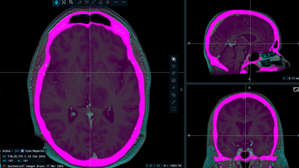
AI overcomes this challenge through generative models—such as Generative Adversarial Networks (GANs) and CycleGAN. Pseudo-planning CT images are synthesized directly from fractional CBCT scans (Spadea et al., 2021). By removing scatter artifacts and restoring standard HU values prior to registration, the process transforms a complex multi-modality problem into a uniform CT-to-CT registration task.
Additionally, deep convolutional neural networks extract multi-scale deep spatial features rather than relying on raw voxel intensities alone, allowing the model to recognize anatomical boundaries regardless of local noise or contrast dropouts (Spadea et al., 2021).
3.2. Resolving Non-Physical Deformations and Mass Discrepancies
Traditional DIR algorithms struggle when anatomical mass is lost (such as tumor shrinkage or weight loss) or when rigid bone structures sit adjacent to deformable soft tissue, resulting in grid folding or unnatural stretching.
AI addresses these physical discrepancies by incorporating mathematical penalties and spatial smoothness losses directly into training (Balakrishnan et al., 2018). This enforces diffeomorphic mapping, ensuring mathematically plausible, continuous deformation vector fields (DVFs) that preserve anatomical topology and prevent unphysical grid tearing or overlapping.
Rather than forcing a pure spatial warp across disappearing tissue volumes, hybrid frameworks combine direct DL segmentation (which naturally handles non-conservative volume changes) with DIR-based propagation.
3.3. Automating Quality Assurance and Speeding Up Workflows
The heavy reliance on manual contour editing and slice-by-slice QA severely limits the efficiency of offline ART workflows.
Conventional iterative registration algorithms can take minutes to solve high-dimensional optimization problems. Trained DL models parameterize registration as a neural network function that computes 3D vector fields in under one second during forward-pass inference (Balakrishnan et al., 2018).
Probabilistic DL frameworks can predict spatial variance maps directly from computed deformation fields (Smolders et al., 2022). By flagging regions with high spatial uncertainty, these models direct clinicians to specific slices requiring manual inspection, eliminating the need to exhaustively review accurate regions.
| Limitation | Conventional DIR Challenge | AI/DL Solution |
|---|---|---|
| CBCT Artifacts | Intensity mismatches throw off gradient matching | Synthetic CT generation & multi-scale deep feature extraction (Spadea et al., 2021) |
| Mass Discrepancies | Unphysical vector stretching / grid folding | Physics-informed loss functions & registration-guided segmentation (Balakrishnan et al., 2018) |
| Labor-Intensive QA | Slow iterative runs & mandatory manual review of all slices | Sub-second inference speeds (Balakrishnan et al., 2018) & DVF uncertainty mapping (Smolders et al., 2022) |
AI is also used to bypass the traditional contour propagation step entirely. Instead of taking the baseline planning CT (pCT) contours and mathematically “stretching” them onto the fractional image via a DVF, AI models (such as 3D U-Nets and Vision Transformers) can perform direct auto-segmentation on the fractional scan.
Contour propagation serves as a fundamental backbone for offline adaptive radiotherapy, bridging the gap between initial treatment planning and ongoing anatomical variations. While traditional deformable image registration techniques have significantly accelerated clinical workflows and improved dosimetric precision, their susceptibility to image artifacts, non-physical deformations, and extensive manual quality assurance limits their potential.
The integration of DL and AI—through synthetic CT generation, physics-informed registration, sub-second inference speeds, and automated uncertainty mapping—effectively addresses these long-standing barriers. As these advanced AI-driven solutions continue to mature and integrate into standard clinical pipelines, offline contour propagation becomes increasingly robust, efficient, and precise, ultimately paving the way toward safer, highly personalized, and fully adaptive radiation oncology care.
References
Balakrishnan, G., Zhao, A., Sabuncu, M. R., Guttag, J., & Dalca, A. V. (2018). VoxelMorph: A Learning Framework for Deformable Medical Image Registration. IEEE Transactions on Medical Imaging, 38(8), 1788–1800. https://doi.org/10.1109/TMI.2019.2897538
Cagni, E., Botti, A., Orlandi, M., Galaverni, M., Iotti, C., Iori, M., Lewis, G., & Spezi, E. (2022). Evaluating the Quality of Patient-Specific Deformable Image Registration in Adaptive Radiotherapy Using a Digitally Enhanced Head and Neck Phantom. Applied Sciences, 12(19), 9493. https://doi.org/10.3390/app12199493
Dona Lemus, O. M., Cao, M., Cai, B., Cummings, M., & Zheng, D. (2024). Adaptive Radiotherapy: Next-Generation Radiotherapy. Cancers, 16(6), 1206. https://doi.org/10.3390/cancers16061206
Gu, X., Dong, B., Wang, J., Yordy, J., Mell, L., Jia, X., & Jiang, S. B. (2013). A contour-guided deformable image registration algorithm for adaptive radiotherapy. Physics in Medicine and Biology, 58(6), 1889–1901. https://doi.org/10.1088/0031-9155/58/6/1889
Hardcastle, N., van Elmpt, W., De Ruysscher, D., Bzdusek, K., & Tomé, W. A. (2013). Accuracy of deformable image registration for contour propagation in adaptive lung radiotherapy. Radiation Oncology, 8, 243. https://doi.org/10.1186/1748-717x-8-243
Li, X., Zhang, Y. Y., Shi, Y. H., Zhou, L. H., & Zhen, X. (2016). Evaluation of deformable image registration for contour propagation between CT and cone-beam CT images in adaptive head and neck radiotherapy. Technology and Health Care, 24, S747–S755. https://doi.org/10.3233/thc-161204
Li, X., Zhang, Y., Shi, Y., Wu, S., Xiao, Y., Gu, X., Zhen, X., & Zhou, L. (2017). Comprehensive evaluation of ten deformable image registration algorithms for contour propagation between CT and cone-beam CT images in adaptive head & neck radiotherapy. PLOS ONE, 12(4), e0175906. https://doi.org/10.1371/journal.pone.0175906
Rong, Y., Rosu-Bubulac, M., Benedict, S. H., Cui, Y., Ruo, R., Connell, T., Kashani, R., Latifi, K., Chen, Q., Geng, H., Sohn, J., & Xiao, Y. (2021). Rigid and Deformable Image Registration for Radiation Therapy: A Self-Study Evaluation Guide for NRG Oncology Clinical Trial Participation. Practical Radiation Oncology, 11(4), 282–298. https://doi.org/10.1016/j.prro.2021.02.007
Smolders, A., Lomax, A., Weber, D. C., & Albertini, F. (2023). Patient-specific neural networks for contour propagation in online adaptive radiotherapy. Physics in Medicine & Biology, 68(9), 095010. https://doi.org/10.1088/1361-6560/accaca
Smolders, A., Lomax, A., Weber, D. C., & Albertini, F. (2022). Deformable Image Registration Uncertainty Quantification Using Deep Learning for Dose Accumulation in Adaptive Proton Therapy. Lecture Notes in Computer Science, 13264, 57–66. https://doi.org/10.1007/978-3-031-11203-4_7
Spadea, M. F., Maspero, M., Zaffino, P., & Seco, J. (2021). Deep learning based synthetic‐CT generation in radiotherapy and PET: A review. Medical Physics, 48(11), 6537–6566. https://doi.org/10.1002/mp.15150