MVision Contour+ is an AI-powered auto-segmentation used in radiation therapy planning. Recently, a series of studies published in peer-reviewed journals evaluated its performance and impact on clinical practice. These studies involved 17 clinics across 12 countries, with 64 evaluators assessing 16,454 volumes of interest on 1,242 scans. None of these studies received financial support from MVision, ensuring unbiased results.’
The research covered a broad range of evaluations, including accuracy, clinical acceptability, time savings, dosimetric impact, and workflow efficiency. This article summarizes the key findings of these studies, organized into five main sections. A list of the published articles with direct links can be found at the end of the article.


Accuracy of Contours
A total of 11 studies assessed the accuracy of the contours produced by MVision Contour+ [1-11]. The results showed consistent, high-quality contours for various anatomical structures. The median DSC for the AI contours in a study including breast cancer cases was 0.87, and another study evaluating multiple anatomic sites reported a similar value, namely 0.88 [5,8].
**The numbers in brackets represent the corresponding number for the studies mentioned in Table 1 (please see below).
Clinical Acceptability
Five studies evaluated the clinical acceptability of MVision Contour+ [3,6,8,11,12]. The tool demonstrated high acceptance among clinicians, with most of the contours requiring only minor edits. The article by Strolin S et al mentions that 87% of the automatically generated contours were considered “well done” or “very well done” by the radiation oncologists involved in the project [3]. In the study by Langmack K et al, 80% of the contours needed no edits or minor edits [12]. In a study comparing multiple AI solutions, MVision achieved the highest percentage (78%) of clinical target volumes that did not need significant corrections, showcasing its reliability in clinical settings [11].
Time Savings
Eight manuscripts reported substantial time savings when using MVision Contour+ [1, 3-7, 9,12]. Across different clinical scenarios, the tool reduced contouring time by a median value ranging from 29% to 79%, and a maximum ranging from 54% to 92% [3]. Another study highlighted that automated segmentation using MVision was significantly faster than manual methods (1.1 minutes vs. 20 minutes) while maintaining contour accuracy [4]. These efficiencies enabled clinicians to allocate more time to other critical tasks, improving workflow and productivity.
Dosimetric Impact
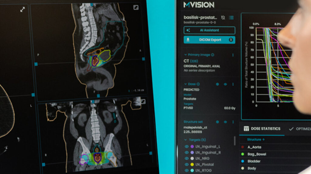
Three studies investigated the dosimetric impact of MVision-generated contours [4,6,12]. The studies indicated that the use of AI-generated contours had minimal impact on radiation dose distribution, suggesting that the automated process could potentially maintain treatment quality and safety [12]. Additionally, the use of MVision Contour+ facilitated the implementation of advanced techniques like volumetric-modulated arc therapy (VMAT), contributing to better dose management [6].
Impact on Workflow
Two articles specifically evaluated the impact on clinical workflow [6,12]. Clinicians experienced increased efficiency, with median planning process turnaround times reduced by up to 9 working days for prostate cancer treatment. The introduction of MVision also allowed oncologists to improve their work-life balance and focus more on professional development and research [12].
Conclusion
Overall, the independent evaluation of MVision Contour+ across a wide range of clinical settings demonstrates its effectiveness in improving the accuracy, efficiency, and workflow of radiation therapy planning. The consistent high-quality performance, combined with significant time savings and minimal impact on dosimetric outcomes, highlights the potential of AI-assisted tools to enhance clinical practice without compromising patient care. These findings support the integration of AI-driven auto-contouring solutions like MVision Contour+ into modern radiotherapy workflows, contributing to better treatment outcomes and improved clinical operations.
The excellent performance reported in these studies was achieved using earlier versions of Contour+. Since then, additional models and structures have been released.
One study involving 97 participants from 23 countries evaluated the role of AI auto-contouring in learning and education [13].
There are other 20 clinical research studies which were presented as posters in international conferences, for which the full text paper is under preparation or in the publication process.
Table 1. Studies published in peer-reviewed journals evaluating MVision Contour+ quality and impact on clinical use