How Adaptive Radiotherapy Addresses Anatomical Changes in Cancer Treatment
The clinical efficacy of conventional radiotherapy is sometimes limited by the morphological variability of organs and anatomical structures over a…
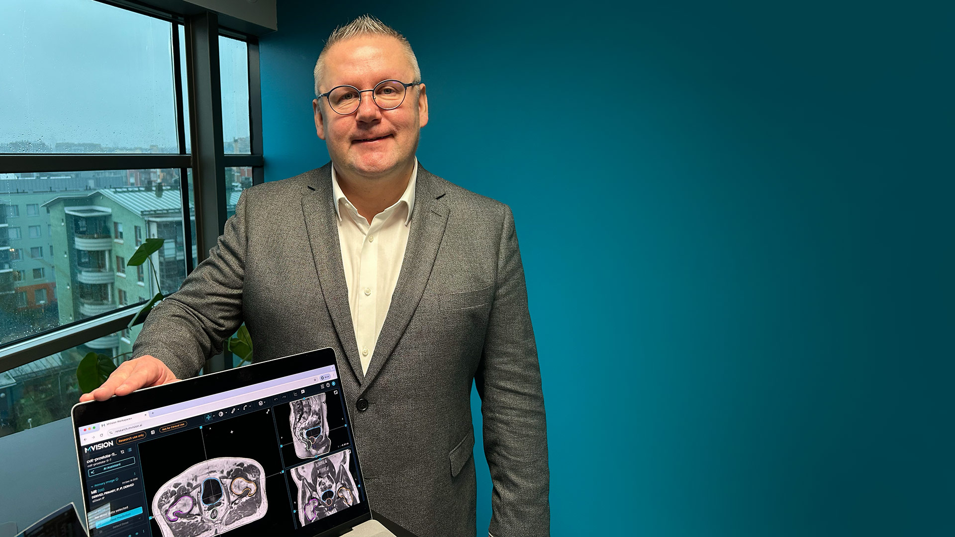
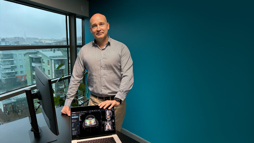
My name is Jani Keyriläinen and I work as a chief physicist (associate professor, PhD) at Turku University Hospital (Tyks) in Turku, Finland.
Turku University Hospital is the second oldest hospital in the Nordic countries and serves a population of about 495,000 people. In radiotherapy, we treat roughly 1,600 patients per year, with about 120 sessions delivered each day.
Our department includes 33 therapists and radiographers, 8 radiation oncologists with 2 in training, 6 medical physicists with 1 in training, as well as nurses, secretaries, and technical support staff.
For external radiotherapy, we operate five linear accelerators, including several Varian TrueBeam and TrueBeam STx units, and a Varian Halcyon 2.0. We deliver a wide range of techniques, such as VMAT, SGRT, SRS, SBRT and TBI. We also have one HDR afterloader for internal radiotherapy.
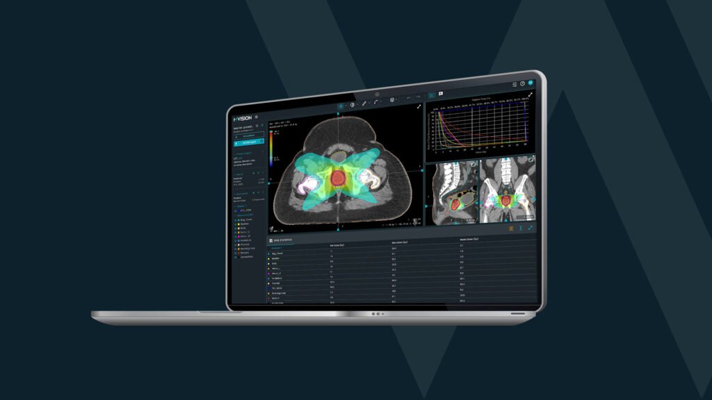
For treatment planning, we use MRI and CT systems tailored for radiotherapy, have access to PET/MRI and PET/CT, and work with Aria, Eclipse and MVision AI Workspace+.
MVision AI Contour+, which is used clinically on almost all of our radiotherapy patients, has been found to be a very effective tool, meaning that compared to manual contouring, it is both accurate versus individual variation and saves contouring time.
For MVision AI products, we were the first university test hospital in 2017–2018. Since 2017, we have had a close research collaboration with MVision AI and the current research collaboration agreement is valid until 2027. It is, of course, possible to continue it also after that.
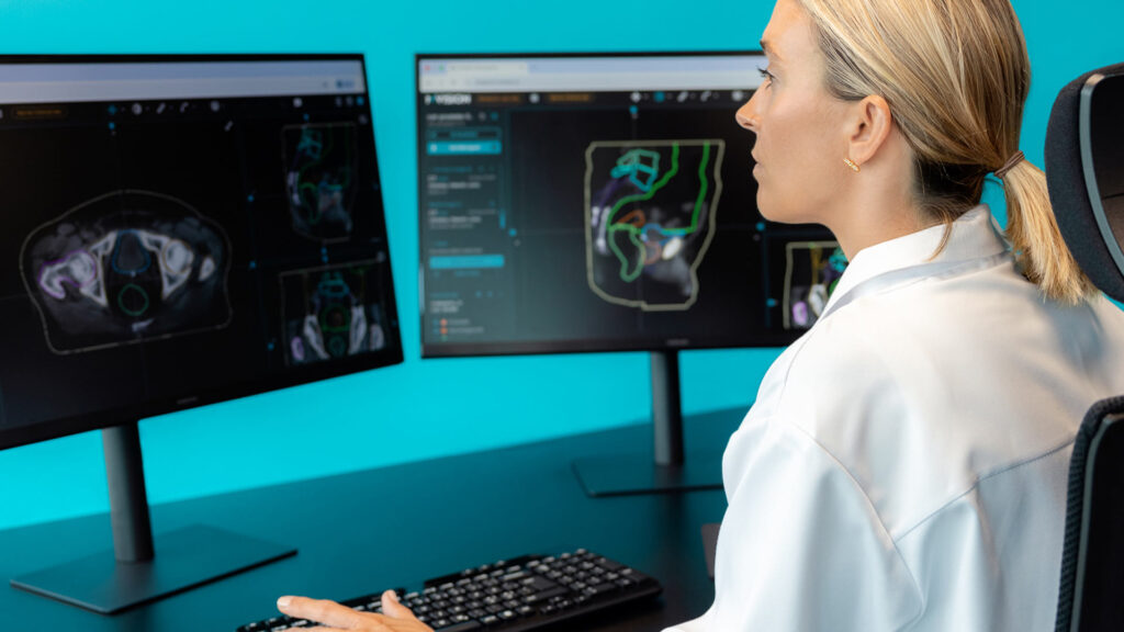
Contour+ does in a few minutes a task that would normally take a radiation oncologist several dozen minutes. Of course, the radiation oncologist still has to monitor the quality of the structures produced by the AI.
The structures determined by the AI are more reliable in the sense that it delineates the structures in a fairly repeatable way, whereas in manual delineation of structures, the result can vary by 5–10 percent by one doctor between different days, not to mention the differences between different doctors.
Using the software frees up doctors’ time for other patient work. Software updates always arrive on time, and there have been very few technical problems.
It is a good thing for us that MVision’s AI developers are located in Finland. Technical support and the implementation of development ideas work agilely and quickly.
The segmentation results are so good that they hardly need to be corrected. It really works automatically and saves a lot of human resources.
The product is based on international guidelines for contouring patient structures in radiotherapy, which was not very common earlier and this philosophy has clearly been an advantage for MVision AI.
MVision AI products are better in quality than other similar software we have used or tested.
Hopefully, in the future, AI will also be used to help with other stages of radiotherapy treatment planning, such as the automatic creation of treatment plans, the analysis of reference images, and quality control.
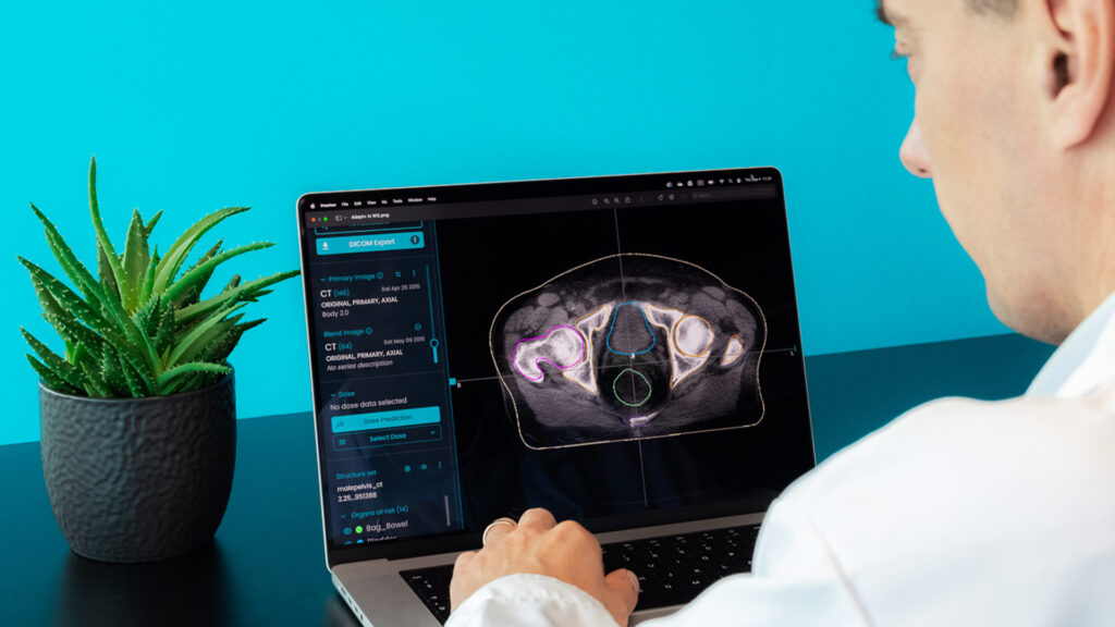
Currently, Contour+ is routinely used clinically. New modules (Image+, Adapt+, Dose+) have been tested to some extent, and the goal is to be able to use them properly in the future. They would make it possible to provide more personalized treatment based on daily CBCT imaging.
At least for now, based on the tests, it seems that they work well. In Tyks, we are actually pioneers for about ten years now in utilizing synthetic CT produced in magnetic resonance imaging, so-called MRI-only based radiotherapy treatment planning. Currently, all of our radiotherapy treatments for pelvic cancers and also some brain cancers are planned based on sCT images, which is still quite rare in the world.
These types of tools are also needed because while the number of patients is increasing, the hospital’s human resources do not want to keep up. In this type of software, the most important thing is quality so that no time is wasted on repairing structures and, on the other hand, consistency, which is often not possible if a human being does the same job. As said above, even with the same doctor, the result can vary by 5–10 percent on two different days.
![]()
About Turku University Hospital
Turku University Hospital (Tyks) provides specialised medical care for the residents of Southwest Finland and also delivers university-level services for the regions of Satakunta and Vaasa. It employs around 8,000 people and offers a wide range of clinical services and training opportunities. Founded in 1756, Tyks is one of the oldest hospitals in Northern Europe and continues to serve as a major academic and clinical centre in Finland.