Recently published data give a glimpse on the time spent for treatment preparation in UK radiation oncology centres. As expected,organ-at-risk and target contouring contouring is the most time-consuming step of the process. However, multiple factors influence the amplitude of this interval, and these new findings can be used as further reference. When artificial intelligence gets widely (and wisely) used, things are changing for the better.
Recent real-world data
Accuracy of contouring is one of the pillars of an adequate treatment plan, when it comes to Radiation Oncology. Fortunately, new technologies have better precision compared with traditional ones, which allows higher dose delivery accuracy by optimising coverage of the target and maximising normal tissue sparing. Increased complexity of fractionation schemes (SIB) and target shapes requires careful delineation of a higher number of organ-at-risk structures, which adds additional pressure on a healthcare system that functions with limited human resources. Moreover, the implementation of modern techniques such as stereotactic irradiation and the shortage of personnel are expected to increase in the future, as well as the number of cancer patients.
In this context, a helping hand is welcomed, and the AI has already started to show its usefulness in the Radiation Oncology field, because of its superior performance compared to previous generation tecnology.
Since the majority of published reports represent the practice of single centres, two national-level surveys were conducted in the UK, in order to assess the time required by routine radiation therapy planning steps, and the usual practice regarding radiotherapy contouring and peer-review process [1].
One of the surveys gathered information on individual consecutive cases, and had 147 responses from 56 separate clinics, and the other one provides data about the usual practice, having 292 participants from 55 cancer centres. Despite its limitations, such as the relatively low representativity of 5 consecutive cancer cases and possible changes existent in practice (questionnaire spread during COVID-19 pandemic), the survey brings relevant information from about 90% of UK cancer treatment providers [1].
Contouring time needs
The UK survey reported a median time per case of 85 minutes. Significant differences were found according to
- Tumour location – shorter time was necessary for breast cancers (43 min) and longer time for head and neck and gynaecological cancers (120 min) (Figure 1);
- Treated volumes – including regional lymph nodes substantially increase the contouring time (Figure 2);
- Technology used – while for 3D conformal radiotherapy plans a median time of 56 minutes was necessary, for IMRT/VMAT or SBRT planning the contouring time increased to 85-90 min.
Paediatric cancers, and especially those treated with protons were the most time consuming, with recorded times beyond 200 minutes.
Clinical oncologists were the main professionals involved in the contouring task, being involved in 87% of cases for the clinical target volume (CTV), but also for the organs at risk (in 74% of cases for delineating and 92% for adjusting them) [1].

Figure 1. Contouring times (minutes) for various anatomical sites, based on results reported by Montague et al (2024), from Royal College of Radiologists Contouring Surveys [1]
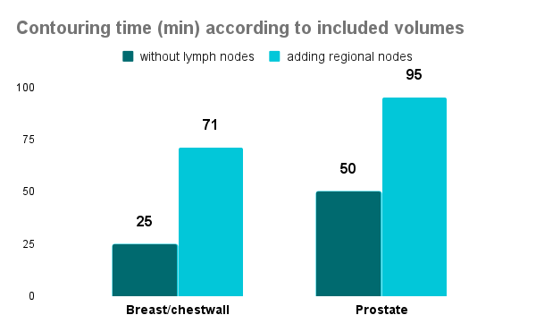
Figure 2. Contouring time (minutes) according to included anatomical structures – data extracted from Montague et al (2024) [1]
Additional time requirements
Except for the contouring task, other steps (such as peer review and approval) imply additional time spent by radiation oncology professionals. Review made by a colleague is increasing the probability of detecting errors in contouring or planning and it was proven to be an effective tool of quality assurance [2,3].
The peer review was performed in less than half of the cases. When it was done, the additional time for peer review was 15 minutes. However, most clinicians did not have any scheduled time in their job plan for peer review [1].
Trainees involvement
Another important aspect was that trainees were involved in the contouring and plan approval process in only 19% of the cases. Almost half of the respondents who worked with trainees reported that their inclusion slowed down the process, about a quarter said it was faster, and the other quarter described no impact on overall time. Overall, trainee involvement added 13 minutes to the planning process [1].
Using of auto-contouring software
Auto-contouring software use was mentioned by 32% of the participants, and most of them for outlining the organs at risk. Adjustments of the automatically generated contours were necessary in most of the situations. However, since the survey took place in 2021, we can assume that the auto-contouring solutions were algorithm and atlas-based, and not using deep-learning technology [1].
Time spent better
The above-mentioned results show in numbers what has been known and felt by numerous radiation oncology professionals. Increasing demands and decreasing workforce create a pressure which can be alleviated by speeding up the process and improving efficiency.
External validation of auto-contouring models provided by MVision AI Contour+ demonstrated significant time savings. Automatic contour generation was usually less than 2 minutes and time needed for reviews and adjustments had a median of 10-13 minutes [4-8]. Taking into account the median contouring time of 85 minutes reported by the survey, subtracting 15 minutes needed to provide final contours based on Contour+ predictions, there are 70 minutes left – enough to perform peer review, train the new generation of healthcare professionals, spend more time with the patient, develop and implement new techniques, read some professional news or take a necessary break, to keep the burnout away.
MVision AI provides also a solution for training in organ-at-risk and lymph nodes contouring, through the new Guide platform. Learning to delineate according to published consensus and guidelines becomes easier, by comparing personal tasks with standardised structures – through visual evaluation and similarity coefficients.
Challenges are real, but also solutions. Contour+ is used across 20 countries in the world, serving more than 120 clinics, and has more than 270 structures, of which 66 lymph node regions, and 20 CTV structures, enabling clinicians to save more time than with organs at risk only. MVision AI was created to offer the best cloud-based technology to support clinicians in their daily practice and continues to develop in the same direction. We are happy to see that our solutions fill a gap in cancer care, enabling radiation oncology professionals across the globe to offer quality treatments, faster.
Reference
- Montague E, Roques T, Spencer K, Burnett A, Lourenco J, Thorp N. How Long Does Contouring Really Take? Results of the Royal College of Radiologists Contouring Surveys. Clin Oncol (R Coll Radiol). Published online March 15, 2024. doi:10.1016/j.clon.2024.03.005
- Ballo MT, Chronowski GM, Schlembach PJ, Bloom ES, Arzu IY, Kuban DA. Prospective peer review quality assurance for outpatient radiation therapy. Pract Radiat Oncol. 2014;4(5):279-284. doi:10.1016/j.prro.2013.11.004
- Qureshi BM, Mansha MA, Karim MU, et al. Impact of Peer Review in the Radiation Treatment Planning Process: Experience of a Tertiary Care University Hospital in Pakistan. J Glob Oncol. 2019;5:1-7. doi:10.1200/JGO.19.00039
- Kiljunen T, Akram S, Niemelä J, et al. A Deep Learning-Based Automated CT Segmentation of Prostate Cancer Anatomy for Radiation Therapy Planning-A Retrospective Multicenter Study. Diagnostics (Basel). 2020;10(11):959. Published 2020 Nov 17. doi:10.3390/diagnostics10110959
- Turcas A, Leucuta D, Balan C, et al. Deep-learning magnetic resonance imaging-based automatic segmentation for organs-at-risk in the brain: Accuracy and impact on dose distribution. Phys Imaging Radiat Oncol. 2023;27:100454. Published 2023 Jun 6. doi:10.1016/j.phro.2023.100454
- Strolin S, Santoro M, Paolani G, et al. How smart is artificial intelligence in organs delineation? Testing a CE and FDA-approved Deep-Learning tool using multiple expert contours delineated on planning CT images. Front Oncol. 2023;13:1089807. Published 2023 Mar 2. doi:10.3389/fonc.2023.1089807
- Bordigoni B et al. Comprehensive Comparison of Four Automatic Tools in OAR Segmentation for Gynaecological Radiotherapy, Physica Medica, Volume 115, November 2023 https://doi.org/10.1016/j.ejmp.2023.102806
- Doolan PJ, Charalambous S, Roussakis Y, et al. A clinical evaluation of the performance of five commercial artificial intelligence contouring systems for radiotherapy. Front Oncol. 2023;13:1213068. Published 2023 Aug 4. doi:10.3389/fonc.2023.1213068