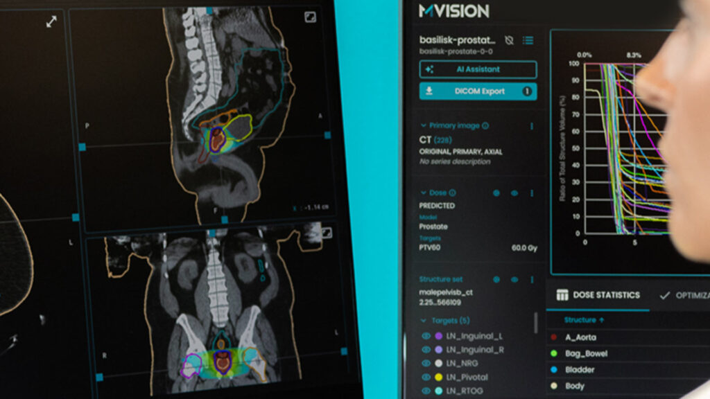
Results of MVision AI’s solutions scientific evaluation were presented at the previous ASTRO annual meeting. One of the posters included data from paired MRI and synthetic CT scans (MRCAT) of male pelvis and showed the accuracy and consistency of Contour+ models. The abstract was published in Volume 117 Issue 2 Supplement of the international Journal of Radiation Oncology *Biology* Physics and can be found here.
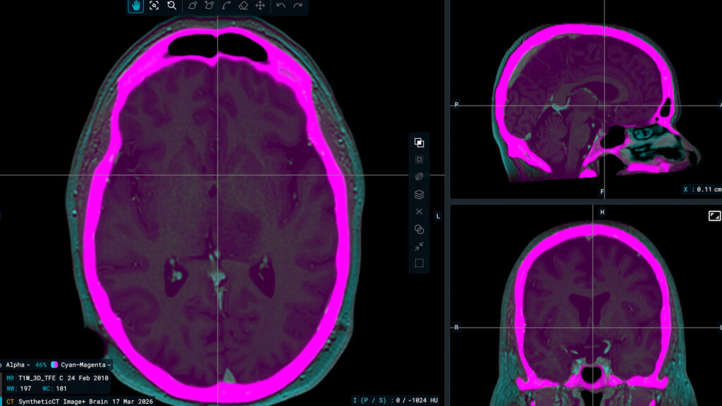
Nowadays MRI scans are used for target delineation and the CTs are needed for planning. However, an MR-only workflow using synthetic CTs (MRCAT) is expected to simplify the process, improve delineation’s accuracy and therefore reduce the risk of systematic errors.
We are adding the full poster content below, with the authors’ approval.
(Text and images copyright retained by the authors)
Clinical Evaluation of Deep Learning-Based Auto-Segmentation toward MRI-Only Prostate Radiotherapy
J.A. Perez-Sanchez 1, R.J. Brisson 1, K.E. Hitchcock 1, R.A. Zlotecki 1, Y. Badyal 2, C. Liu 1, G. Yan 1
1 Department of Radiation Oncology, University of Florida College of Medicine, Gainesville, FL
2 MVision AI, Helsinki, Finland
Purpose/Objective(s)
Due to its superior soft-tissue contrast, MRI is the imaging modality of choice for target and organs-at-risk delineation in prostate radiotherapy. A recent advanced MR scanner allows for an MRI-only workflow owing to its ability to generate synthetic CT (MRCAT) that enables accurate dose calculation and treatment planning. While auto-segmentation algorithms for CT images are increasingly being adopted in clinical practice, there are fewer such algorithms for MRI images. The purpose of this study was to evaluate a deep learning-based auto-segmentation tool (from MVision AI) for both MRI and MRCAT images. We hypothesize that such algorithms could produce accurate contours that require little manual editing, resulting in significant time saving and workflow efficiency in MRI-only treatment planning.
Materials/Methods
T2 MRI and MRCAT image pairs (acquired on a Philips Ingenia 1.5T MRI Scanner) for 10 prostate patients treated in an MRI-only workflow at our institution were retrospectively collected with IRB approval. These image volumes were then segmented using deep learning-based auto-segmentation models trained for MRI and CT images, respectively. The structures were manually edited by an experienced physician in a commercial imaging informatics system (Velocity). Finally, the auto-segmented and the post-edit structures were compared using Dice Similarity Coefficient (DSC) and 95% Hausdorff Distance (HD95).
Results
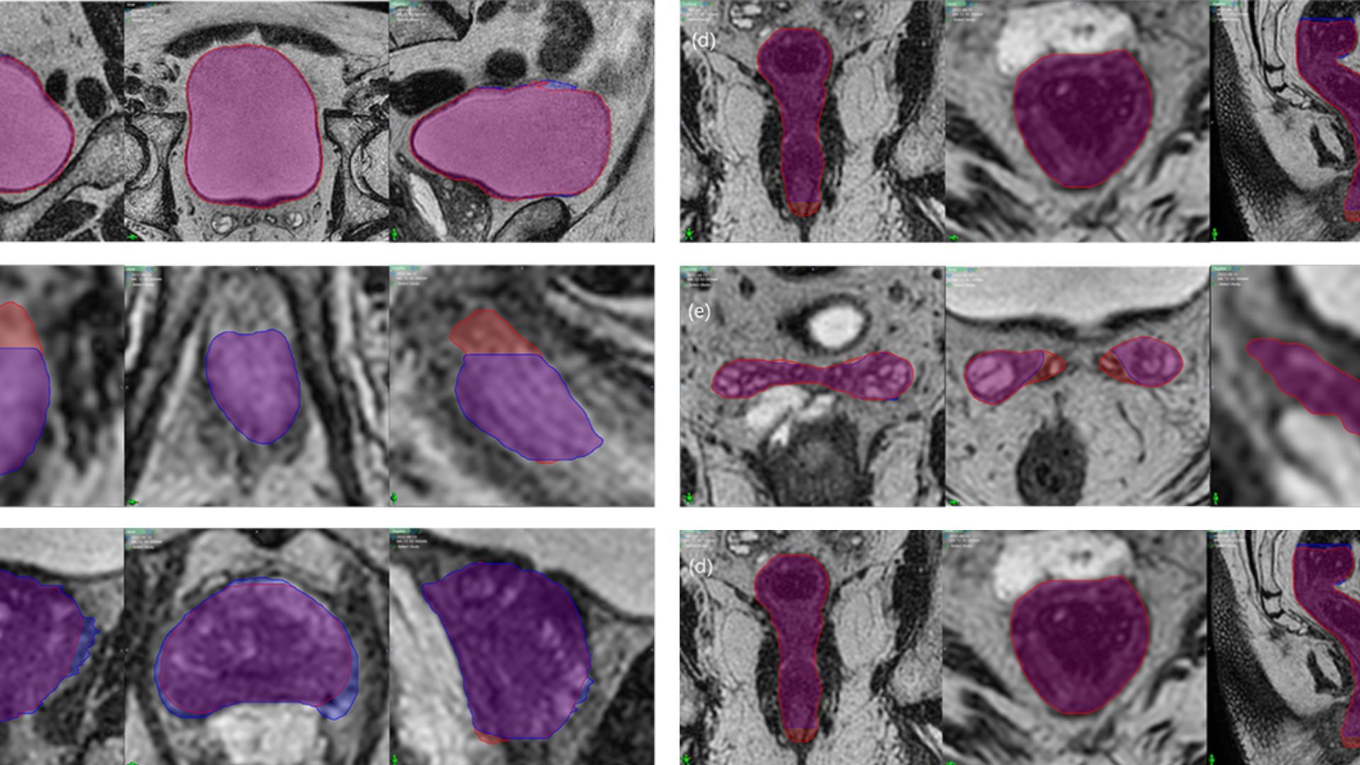
Excellent agreement was found between the auto-segmented and post-edit structures. For MRCAT, the average DSC was 0.92±0.03, 0.99±0.00, 0.90±0.06, 0.89±0.07, 0.89±0.06, 0.95±0.04, and 0.96±0.03 for prostate, bladder, rectum, penile bulb, seminal vesicles, left femur, and right femur, respectively. The average HD95 for these structures were 0.65±0.51mm, 0.01±0.01mm, 2.07±1.68mm, 1.10±0.91mm, 1.67±1.81mm, 0.86±1.24mm, and 0.67±1.49mm, respectively. For MRI, the average DSC was 0.96±0.02, 0.99 ±0.00, 0.96±0.03, 0.87±0.03, 0.94±0.06, and 0.89±0.11 for prostate, bladder, rectum, penile bulb, seminal vesicles, and Barrigel spacer, respectively. The average HD95 was 0.25±0.21mm, 0.00±0.00mm, 1.60±1.87mm, 1.43±0.98mm, 0.50±0.86mm, and 1.56±2.26mm, respectively.

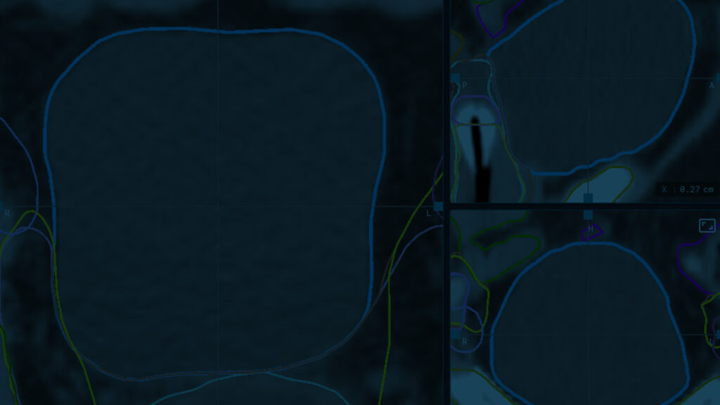
Figure 1. MVision segmentations (red) vs. physician-edited segmentations (blue) on MR images of Patient 1. (a) bladder, (b) penile bulb, (c) prostate, (d) rectum, (e) seminal vesicles. Left-to-right: coronal, axial, sagittal cross-sections.

Figure 2. MVision segmentations (red) vs. physician-edited segmentations (blue) on MRCAT images of patient 3. (a) bladder, (b) penile bulb, (c) prostate, (d) rectum, and (e) seminal vesicles. Left-to-right: coronal, axial, sagittal cross-sections.


Conclusion
We evaluated the clinical acceptability of deep learning-based auto-segmentation models for prostate radiotherapy, one for MRI and one for MRCAT delineation. Both models demonstrated superior accuracy, and a high degree of consistency, offering the potential for significant time saving in treatment planning and improved workflow efficiency.
These excellent results are in line with previous studies and external validation of Contour+ quality The novelty of this study is the confirmation of MVision AI male pelvis models performance on synthetic CTs and opens up new opportunities for using it in the MR-only workflow for treating prostate cancer.